SPHENOID BONE
Interactive 3D Version Available
Explore this anatomy in full 360° rotation with our interactive 3D viewer
View in 3D →SPHENOID BONE AR ATLAS
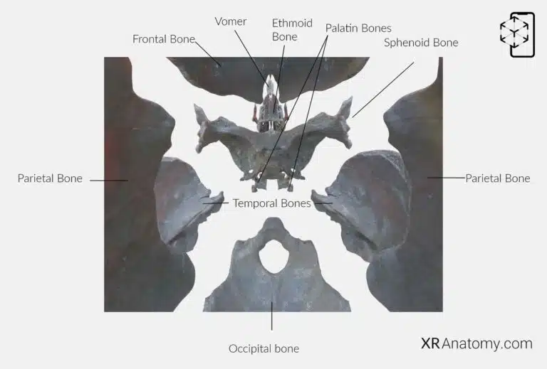
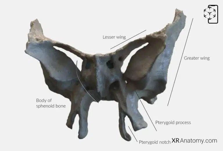
AR Figure 30 – Sphenoid Bone: Disarticulated view, Augmented Illustration by B. Leahu – MD. This image is licensed under the Creative Commons Attribution-NonCommercial-NoDerivs 3.0 Unported (CC BY-NC-ND 3.0).
The is a complex bone situated at the base of the skull, playing a crucial role in the cranial structure. It articulates with numerous bones, including the vomer, ethmoid, frontal, occipital, parietal, temporal, zygomatic, and palatine bones, forming a central wedge that unites the cranial and facial skeletons. Structurally, the sphenoid bone consists of a central , two , two , and two , each contributing to various functions and articulations within the skull.
BODY OF SPHENOID BONE
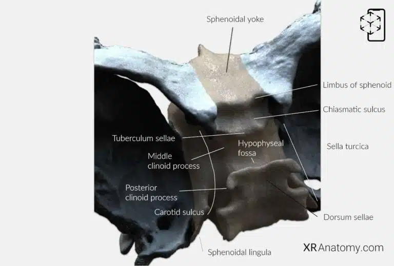
AR Figure 32 – Sphenoid Bone: Body of sphenoid bone, Augmented Illustration by B. Leahu – MD. This image is licensed under the Creative Commons Attribution-NonCommercial-NoDerivs 3.0 Unported (CC BY-NC-ND 3.0).
The is a hollow, cuboid-shaped structure that forms the central portion of the bone. On its superior surface lies the , an elevated area along the midline where the lesser wings converge. Posteriorly, the yoke is bounded by the , beyond which lies the —a horizontal groove accommodating the optic chiasm. Posterior to this sulcus is the , a saddle-shaped depression that houses the pituitary gland within its deepest part, known as the . The forms the anterior boundary of the sella turcica, flanked by the on either side. The constitutes the posterior wall of the sella turcica and bears the at its upper corners, serving as attachment points for dural structures. Laterally, the runs along the sides of the body, forming a broad groove that lodges the internal carotid artery and the cavernous sinus. Adjacent to this groove is the , a bony ridge marking the lateral margin of the carotid sulcus.
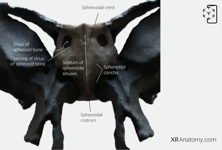
AR Figure 33 – Sphenoid Bone: Body of sphenoid bone, Augmented Illustration by B. Leahu – MD. This image is licensed under the Creative Commons Attribution-NonCommercial-NoDerivs 3.0 Unported (CC BY-NC-ND 3.0).
The anterior surface of the sphenoid body features the , a vertical bony ridge along the midline that articulates with the perpendicular plate of the ethmoid bone. Extending inferiorly from the crest is the , a spine that articulates with the vomer. Within the body lie the —two irregular cavities separated by a thin bony . Each sinus opens into the nasal cavity through openings located on either side of the sphenoidal crest. Covering these openings are the , two thin, curved bony plates that form part of the nasal cavity's roof and contribute to the sinus walls.
LESSER WING
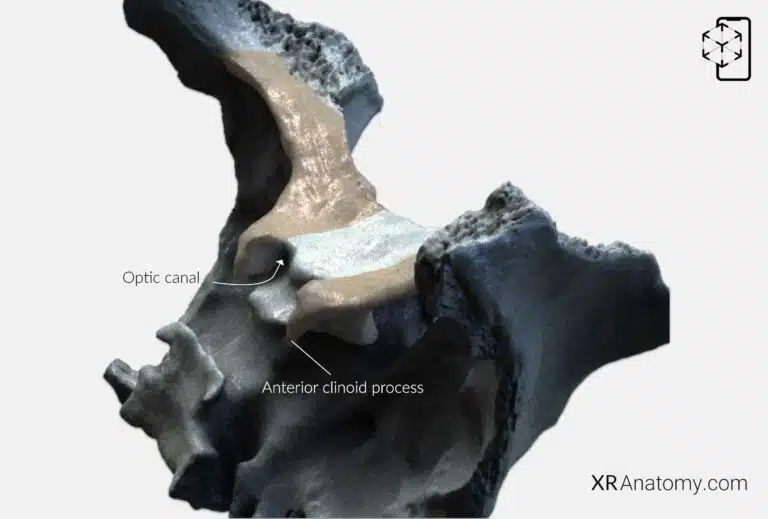
AR Figure 34 – Sphenoid Bone: Lesser wing, Augmented Illustration by B. Leahu – MD. This image is licensed under the Creative Commons Attribution-NonCommercial-NoDerivs 3.0 Unported (CC BY-NC-ND 3.0).
The are two thin, triangular plates that extend laterally from the superior anterior part of the sphenoid body. They form part of the floor of the anterior cranial fossa and the posterior part of the roof of the orbits. The are located at the base of the lesser wings, each serving as a passage for the optic nerve and ophthalmic artery into the orbit. Medially, the posterior borders of the lesser wings give rise to the , which serve as attachment points for the tentorium cerebelli, a fold of dura mater that separates the cerebrum from the cerebellum.
GREATER WING
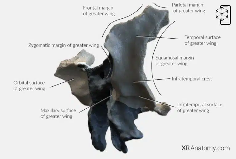
AR Figure 36 – Sphenoid Bone: Greater wing, Augmented Illustration by B. Leahu – MD. This image is licensed under the Creative Commons Attribution-NonCommercial-NoDerivs 3.0 Unported (CC BY-NC-ND 3.0).
The are two strong, curved extensions from the lateral sides of the sphenoid body. They contribute to the formation of the middle cranial fossa, the lateral wall of the skull, and the orbits. The lateral surface of each greater wing is divided by the into the superior and the inferior . The temporal surface forms part of the temporal fossa, while the infratemporal surface contributes to the infratemporal fossa and contains important foramina such as the and . The orbital surface is smooth and forms part of the lateral wall of the orbit. The margins of the greater wing articulate with neighboring bones: the with the zygomatic bone, the with the frontal bone, the with the parietal bone, and the with the temporal bone.
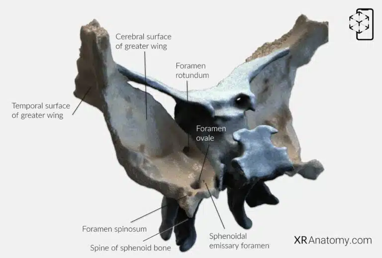
AR Figure 35 – Sphenoid Bone: Greater wing, Augmented Illustration by B. Leahu – MD. This image is licensed under the Creative Commons Attribution-NonCommercial-NoDerivs 3.0 Unported (CC BY-NC-ND 3.0).
The superior () of the greater wing forms part of the middle cranial fossa and features several important openings. The transmits the maxillary nerve, the allows passage of the mandibular nerve and accessory meningeal artery, and the transmits the middle meningeal artery. Occasionally, the and foramen petrosum are present, transmitting emissary veins and the lesser petrosal nerve, respectively. Lateral to the foramen spinosum is the , serving as an attachment for ligaments. The sulcus of the auditory tube runs along the lateral part of the posterior margin, providing attachment for the cartilaginous part of the auditory tube.
PTERYGOID PROCESS
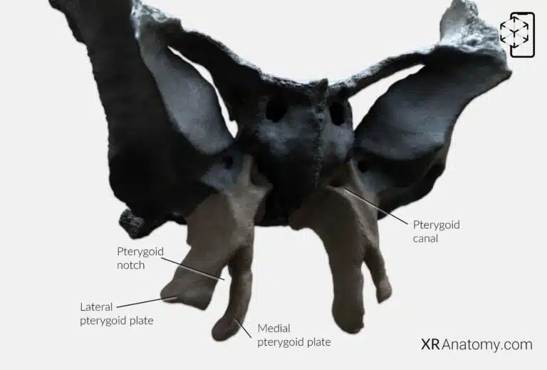
AR Figure 37 – Sphenoid Bone: Pterygoid process, Augmented Illustration by B. Leahu – MD. This image is licensed under the Creative Commons Attribution-NonCommercial-NoDerivs 3.0 Unported (CC BY-NC-ND 3.0).
The extend downward from the junction of the sphenoid body and greater wings. Each process consists of a and a , fused anteriorly but separated inferiorly by the . The lateral pterygoid plate provides attachment for the lateral pterygoid muscle on its lateral surface and the medial pterygoid muscle on its medial surface, both important in mastication. The medial pterygoid plate contributes to the nasal cavity and ends inferiorly in the , a hook-like process that acts as a pulley for the tensor veli palatini muscle.
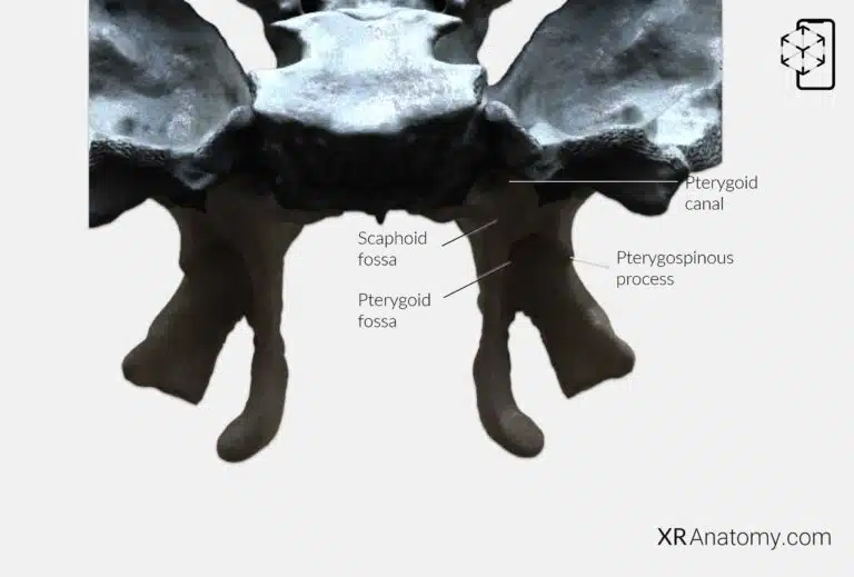
AR Figure 38 – Sphenoid Bone: Pterygoid process, Augmented Illustration by B. Leahu – MD. This image is licensed under the Creative Commons Attribution-NonCommercial-NoDerivs 3.0 Unported (CC BY-NC-ND 3.0).
Between the medial and lateral plates lies the , accommodating muscles of the soft palate. Above this fossa is the , serving as an attachment site for the tensor veli palatini muscle. The , a pointed projection on the posterior border of the lateral pterygoid plate, can occasionally ossify to form a ligament. The passes through the base of the pterygoid process, transmitting the nerve and artery of the pterygoid canal.
VAGINAL PROCESS OF THE SPHENOID BONE
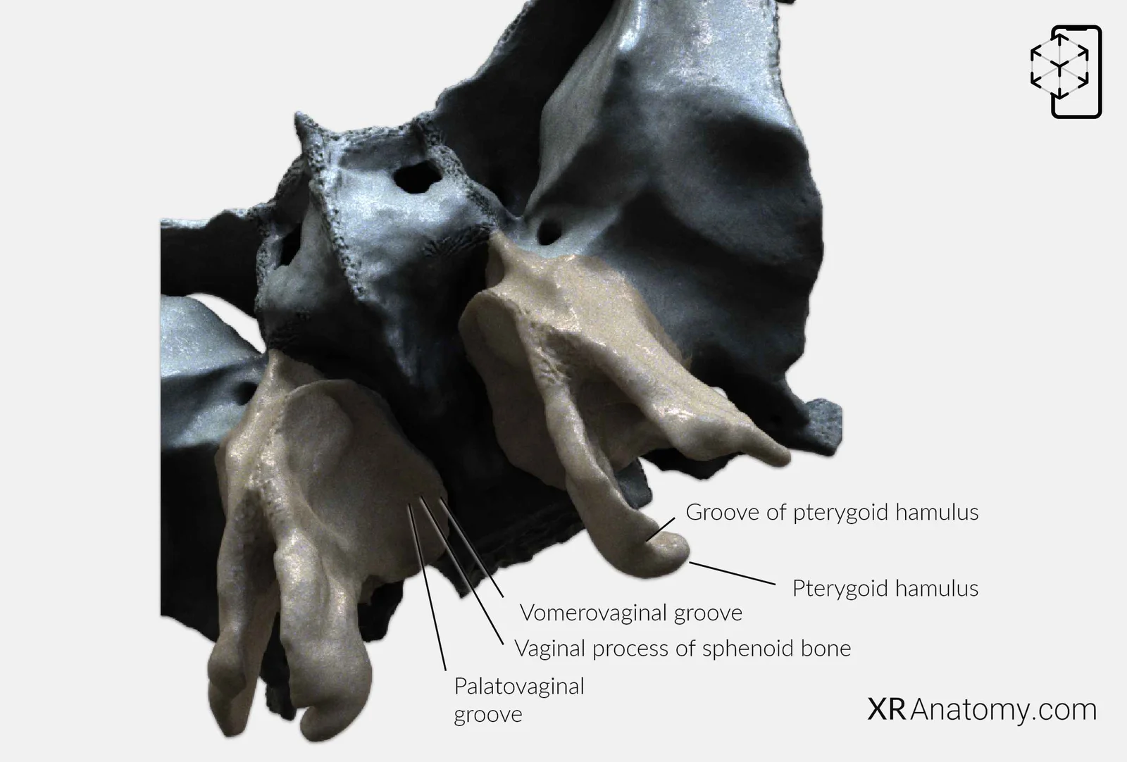
AR Figure 39 – Sphenoid Bone: Vaginal process, Augmented Illustration by B. Leahu – MD. This image is licensed under the Creative Commons Attribution-NonCommercial-NoDerivs 3.0 Unported (CC BY-NC-ND 3.0).
The are small bony projections on each side of the , articulating with the sphenoidal process of the palatine bone and the ala of the vomer. The on the sphenoid bone combines with the palatine bone to form the palatovaginal canal, transmitting the pharyngeal nerve and vessels. Similarly, the forms the vomerovaginal canal when joined by the vomer bone. The , located at the inferior end of the medial pterygoid plate, features a groove guiding the tendon of the tensor veli palatini muscle. The traverses the base of the pterygoid process, with anterior and posterior openings, transmitting the nerve and artery of the pterygoid canal.
BIBLIOGRAPHY
1. Henry G, Warren HL. Osteology. In: Anatomy of the Human Body. 20th ed. Philadelphia: Lea & Febiger; 1918. p. 129–97.
2. Sampson HW, Montgomery JL, Henryson GL. Atlas of the human skull. College Station: Texas A & M University Press; 2007.
4. Saylam C, Özer MA, Ozek C, Gurler T. Anatomical Variations of the Frontal and Supraorbital Transcranial Passages. Journal of Craniofacial Surgery. 2003;14(1):10–2.
5. Hosemann W, Gross R, Goede U, Kuehnel T. Clinical anatomy of the nasal process of the frontal bone (spina nasalis interna). Otolaryngology – Head and Neck Surgery. 2001;125(1):60–5.
6. Steele DG, Bramblett CA. The anatomy and biology of human skeleton. Texas A&M University Press; 1988.
7. Tersigni-Tarrant MTA, Shirley NR. Human osteology. Vol. 4, Forensic Anthropology: An Introduction. 2012. 33–68 p.
8. Monjas-Cánovas I, García-Garrigós E, Arenas-Jiménez JJ, Abarca-Olivas J, Sánchez-Del Campo F, Gras-Albert JR. Radiological Anatomy of the Ethmoidal Arteries: CT Cadaver Study. Acta Otorrinolaringologica (English Edition). 2011;62(5):367–74.
9. Pereira G, Lopes P, Santos A, Krebs. Morphometric aspects of the jugular foramen in dry skulls of adult individuals in Southern Brazil. Vol. 27, J. Morphol. Sci. 2010.
12. Kunc V, Fabik J, Kubickova B, Kachlik D. Vermian fossa or median occipital fossa revisited: Prevalence and clinical anatomy. Annals of Anatomy. 2020 May 1;229:151458.
14. Standring S. The skull. In: Gray’s anatomy: the anatomical basis of clinical practice. 2021st ed. Elsevier Health Sciences; 2021. p. 558–73.
15. Rhoton AL. Chapter 1 Overview of Temporal Bone. Neurosurgery. 2007;
17. Tóth M, Moser G, Patonay L, Oláh I. Development of the anterior chordal canal. Annals of Anatomy. 2006;
18. Carpenter G, Knipe H. Tympanic part of temporal bone. Radiopaedia.org. 2014 Mar 23;
19. Eckerdal O. The petrotympanic fissure: A link connecting the tympanic cavity and the temporomandibular joint. Cranio – Journal of Craniomandibular Practice. 1991;9(1):15–22.
21. Singh R, Kishore Gupta N, Kumar R. Morphometry and Morphology of Foramen Petrosum in Indian Population. Basic Sciences of Medicine. 2020;2020(1):8–9.
23. Piagkou M, Xanthos T, Anagnostopoulou S, Demesticha T, Kotsiomitis E, Piagkos G, et al. Anatomical variation and morphology in the position of the palatine foramina in adult human skulls from Greece. Journal of Cranio-Maxillofacial Surgery. 2012 Oct 1;40(7):e206–10.
