INFERIOR NASAL CONCHA
TABLE OF CONTENTS
Interactive 3D Version Available
Explore this anatomy in full 360° rotation with our interactive 3D viewer
View in 3D →INFERIOR NASAL CONCHA AR ATLAS

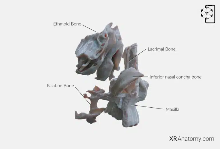
The is a scroll-shaped bone situated on the lateral wall of the nasal cavity. It plays a significant role in the respiratory system by increasing the surface area of the nasal cavity, which helps in warming and humidifying the inhaled air. The bone articulates with several surrounding bones, contributing to the architecture of the nasal cavity. The bones it are: The lacrimal bone, The ethmoid bone, The palatine bone, The maxilla.

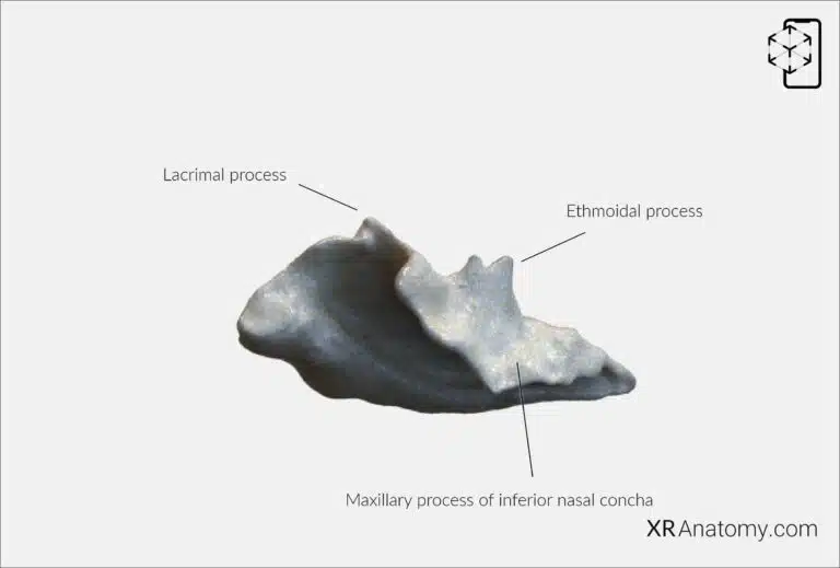
Several processes extend from the inferior nasal concha, each articulating with adjacent bones and contributing to specific functions. The maxillary process of the inferior nasal concha extends laterally to articulate with the maxilla. This process helps form part of the maxillary sinus wall, further integrating the inferior nasal concha into the facial skeleton and supporting the sinus structure.
BIBLIOGRAPHY
1. Henry G, Warren HL. Osteology. In: Anatomy of the Human Body. 20th ed. Philadelphia: Lea & Febiger; 1918. p. 129–97.
2. Sampson HW, Montgomery JL, Henryson GL. Atlas of the human skull. College Station: Texas A & M University Press; 2007.
4. Saylam C, Özer MA, Ozek C, Gurler T. Anatomical Variations of the Frontal and Supraorbital Transcranial Passages. Journal of Craniofacial Surgery. 2003;14(1):10–2.
5. Hosemann W, Gross R, Goede U, Kuehnel T. Clinical anatomy of the nasal process of the frontal bone (spina nasalis interna). Otolaryngology – Head and Neck Surgery. 2001;125(1):60–5.
6. Steele DG, Bramblett CA. The anatomy and biology of human skeleton. Texas A&M University Press; 1988.
7. Tersigni-Tarrant MTA, Shirley NR. Human osteology. Vol. 4, Forensic Anthropology: An Introduction. 2012. 33–68 p.
8. Monjas-Cánovas I, García-Garrigós E, Arenas-Jiménez JJ, Abarca-Olivas J, Sánchez-Del Campo F, Gras-Albert JR. Radiological Anatomy of the Ethmoidal Arteries: CT Cadaver Study. Acta Otorrinolaringologica (English Edition). 2011;62(5):367–74.
9. Pereira G, Lopes P, Santos A, Krebs. Morphometric aspects of the jugular foramen in dry skulls of adult individuals in Southern Brazil. Vol. 27, J. Morphol. Sci. 2010.
12. Kunc V, Fabik J, Kubickova B, Kachlik D. Vermian fossa or median occipital fossa revisited: Prevalence and clinical anatomy. Annals of Anatomy. 2020 May 1;229:151458.
14. Standring S. The skull. In: Gray’s anatomy: the anatomical basis of clinical practice. 2021st ed. Elsevier Health Sciences; 2021. p. 558–73.
15. Rhoton AL. Chapter 1 Overview of Temporal Bone. Neurosurgery. 2007;
17. Tóth M, Moser G, Patonay L, Oláh I. Development of the anterior chordal canal. Annals of Anatomy. 2006;
18. Carpenter G, Knipe H. Tympanic part of temporal bone. Radiopaedia.org. 2014 Mar 23;
19. Eckerdal O. The petrotympanic fissure: A link connecting the tympanic cavity and the temporomandibular joint. Cranio – Journal of Craniomandibular Practice. 1991;9(1):15–22.
21. Singh R, Kishore Gupta N, Kumar R. Morphometry and Morphology of Foramen Petrosum in Indian Population. Basic Sciences of Medicine. 2020;2020(1):8–9.
23. Piagkou M, Xanthos T, Anagnostopoulou S, Demesticha T, Kotsiomitis E, Piagkos G, et al. Anatomical variation and morphology in the position of the palatine foramina in adult human skulls from Greece. Journal of Cranio-Maxillofacial Surgery. 2012 Oct 1;40(7):e206–10.

